Camera manufacturer Nikon has been running the International Small World Competition since 1975. Now over 40 years old, the competition celebrates the very best in photomicrography.
A photomicrograph is a digital image taken through a microscope to magnify an object several times to show close up features at a scale visible for us to see.
Anyone, anywhere can enter the Nikon Small World competition, with the subject matter left entirely open so participants can choose to take photos of whatever they wish. A panel of expert judges looks over all the entries before deciding upon 20 winners each year.
We're taking a look back at all the first prize winners dating back to the competition's inception in 1975.
- 53 stunning photos from The Nature Conservancy global photo contests
- 57 stunning photos from the National Sony World Photography Awards
- 37 amazing photos from the Astronomy Photographer of the Year awards
1975: James Dvorak, USA

- Subject Matter: Oxalic acid crystals during Precipitation
- Magnification: 100x
- Technique: Transmitted Polarized Light with a Berek Quartz Wedge
James Dvoark has employed the use of polarised light in this image. Polarised light is formed by passing light through a polarising filter to transmit light in just one direction. Microscopes have two polarising filters, one above and one below the sample, these are called the polariser and the analyser respectively. Polarised light can help reveal the structure and composition of samples, which is clearly shown in James' image.
1976: Eric V. Gravé, New York, USA

- Subject Matter: Encysted Parasitic round worm (trichinella spirals)
- Magnification: 50x
- Technique: Differential Interference Contrast
Differential Interference Contrast (DIC) produces contrast by visually displaying the refractive index gradients of different areas of a specimen. It's a relatively complex microscopy method, developed by Georges Nomarski in 1952.
James W. Smith, Independence, Ohio, USA

The 1977 winner of the Small World Competition used DIC to produce this image of a 305 times magnified look at Crystals of rutile and tridymite.
- Subject Matter: Crystals of rutile (titanium dioxide) and tridymite (a polymorph of quartz) in a cobalt-rich glass
- Magnification: 350x
- Technique: Nomarski Differential Interference Contrast
The 1977 winner of the Small World Competition also used DIC to produce the final image. The crystals in the image wouldn't have been so colourful to the naked eye, so the bright colours have been produced using the DIC method.
1978: David Gnizak, Independence, Ohio, USA

This one shows vaporised gold shown at 55 times magnification. An impressive view of a precious metal.
- Subject Matter: Gold, vaporized in a tungsten boat, in a vacuum evaporator
- Magnification: 55x
- Technique: Nomarski Differential Interference Contrast
DIC must have become a popular method for photomicrography fans as it produced yet another winner in 1978.
1979: Paul W. Johnson, University of Rhode Island, Kingston, Rhode Island, USA

This curious view shows a atalked protozoan attached to a filamentous green algae with bacteria on its surface.
- Subject Matter: Stalked protozoan attached to a filamentous green algae with bacteria on its surface
- Magnification: 160x
- Technique: Nomarski Differential Interference Contrast
1979 would see the last of a solid winning run for the DIC method, but only until 1981.
1980: James M. King, UC Santa Barbara, Marine Science Institute, Santa Barbara, California, USA

- Subject Matter: Larvacean within its feeding structure dyed with red organic carmine which the larvacean syphoned in while filter feeding
- Magnification: 20x
- Technique: Underwater camera with multiple extension tubes
An extension tube is a component that fits between the camera and the lens, with the purpose of moving the lens away from the camera's internal sensor. It provides a simple and cheaper way of getting closer to the subject without paying for an expensive macro lens.
1981: David Gnizak, Ferro Corporation Research Center, Independence, Ohio, USA

David Gnizak produced this incredible award-winning image of collapsed bubbles from an annealed experimental electronic sealing glass back in 1981.
- Subject Matter: Collapsed bubbles from an annealed experimental electronic sealing glass
- Magnification: 55x
- Technique: Reflected Light, Nomarski Differential Interference Contrast
We promised another Differential Interface Contrast photo winner in 1981 and here it is. This example by now multiple award winner David Gnizak is yet more proof that photomicrography can produce stunning works of art.
1982: Dr. Jonathan Eisenback, North Carolina State University, Department of Plant Pathology, Raleigh, North Carolina, USA

- Subject Matter:: Silverberry scaly hair whole mount
- Magnification: 400x
- Technique:: Brightfield
Brightfield is seen as the simplest form of photomicrography and involves light being passed through, or in some cases reflected off a specimen. This image shows a Silver berry scaly hair flowering plant, magnified 400 times.
1983: Elieen Roux, Bob Hope International Heart Research Institute, Seattle, Washington, USA

Darkfield microscopy is a method used to create contrast in unstained samples. The resulting images have a bright sample and an incredibly dark background.
- Subject Matter: Suctorian attached to stalk of red algae, encircled by ring of diatoms
- Magnification: 125x
- Technique: Darkfield
Darkfield microscopy is a method used to create contrast in unstained samples. Resulting images have a bright sample and an incredibly dark background.
1984: John I. Koivula, Gemological Institute of America, Carlsbad, California, USA

- Subject Matter: Inclusions of goethite and hematite in Brazilian agate
- Magnification: 30x
- Technique: Transmitted Light with Reflected Fiber-optic Illumination
While not specified for this image, a fiber-optic ring light is the most widely used form of finer-optic illumination. It's fixed in position and surrounds the entire microscope, meaning it can provide a consistent quality of light onto the sample.
1985: Dr. Jonathan Eisenback, North Carolina State University, Department of Plant Pathology, Raleigh, North Carolina, USA

- Subject Matter: Formalin-fixed whole mount of a spiral nematode, multiple exposure
- Magnification: 160x
- Technique: Darkfield
This image is another great example of dark field photography, clearly showing a bright-coloured sample with a dark background. This particular image is of a spiral nematode, one of the most common parasitic nematodes of plants, found in corn, bananas and soy beans.
1986: Dr. Stephen Lowry, University of Ulster, Department of Biology, Coleraine, Northern Ireland, United Kingdom

Here a type of fresh-water organism is seen capturing a Daphina pulex, the most common species of water flea, found in the America, Europe and Australia.
- Subject Matter: Live water mount of Hydra viridissima capturing Daphnia pulex
- Magnification: 10x
- Technique: Darkfield
If you've been following closely, you'll know that this photo is another example of darkfield microscopy. In this image a Hydra viridissima (green hydra) a type of fresh-water organism is seen capturing Daphina pulex, the most common species of water flea, found in the America, Europe and Australia.
1987: Julie Macklin & Dr. Graeme Laver, Australian National University, Canberra, Australia

Here's a curious 14x magnified view of viral neuraminidase. Found on the surface of the influenza virus,
- Subject Matter: Crystals of influenza virus neuraminidase isolated from terns
- Magnification: 14x
- Technique: Brightfield with Coloured Filters
Another example of a brightfieqld image won the Small World competition in 1987. This example shows a 14x magnified view of viral neuraminidase, which is found on the surface of the influenza virus and that allows the virus to be released from its host cells.
1988: David A. Smith Victoria Point, Queensland, Australia

- Subject Matter: Gold residue and gold-coated bubbles in glassy matrix
- Magnification: 20x
- Technique: Brightfield
Another example of brightfieqld imaging shows just how common and popular it is, while also proving it can produce stunning images. This image doesn't use living organisms or cells for its subject, but gold instead. The result looks very much like a painting, and we love it.
1989: Marc Van Hove, Centexbel, Zwijnaarde, Belgium

- Subject Matter: Multiple exposure of a knitting machine needle
- Magnification: 10x
- Technique: Brightfield
- We bet if you tried guessing what a knitting machine needle would look like when put under a microscope you'd never imagine it would produce an image like this. If we weren't told what the subject was, it probably would have taken us some time to realise what it was.
1990: Richard H. Lee, Argonne National Laboratory, Argonne, Illinois, USA

No, that's not a fish that's been put under the microscope, just polarised light reacting to the crystals.
- Subject Matter: Crystals evaporated from solution of magnesium sulfate and tartaric acid
- Magnification: 50x
- Technique: Polarised Light
No, that's not a fish that's been put under the microscope, but the way the in which the polarised light has reacted to the crystals has left a rather serene image.
1991: Marc Van Hove, Centexbel, Zwijnaarde, Belgium

1991's winner showed a a 25x magnified image of an elastic fiber; a simple subject and a simple method, but a very artistic image as a result.
- Subject Matter: Polyurethane elastic fiber bundle
- Magnification: 25x
- Technique: Polarised Light
Another win for Marc Van Hove in 1991. This time he's taken a 25x magnified image of an elastic fiber; a simple subject and a simple method, but a very artistic image as a result.
1992: Lars Bech, Deurne, The Netherlands

This one shows a 35 times magnified view of a 10-year old preparation of barbital, fenacetine, valium and acetic acid
- Subject Matter: 10-year old preparation of barbital, fenacetine, valium and acetic acid
- Magnification: 35x
- Technique: Polarised Light
Lars Bech of the Netherlands has taken a 35x magnified image of a mixture of three different drugs with acetic acid, which is the main ingredient of vinegar other than water.
1993: Ron Sturm, Construction Technology Laboratories, Inc. Skokie, Illinois, USA

- Subject Matter: Fossil Fusulinids in limestone
- Magnification: 8x
- Technique: Polarised Light
Another example of a photomicrograph taken using the polarising light method comes from Ron Sturm in 1993. This image shows a couple of fossils in limestone and is only magnified 8x. The human eye would likely be able to see the shape of the fossils, but the polarised light has revealed the markings.
1994: Jean Rüegger-Deschenaux, Mikroskopische Gesellschaft, Zurich, Switzerland

- Subject Matter: Cross-section of very young beech
- Magnification: 40x
- Technique: Brightfield
This brightfield photograph of a young beech tree has produced a fascinating image that looks a little bit like a grapefruit. We'd be interested to see what a much older beech tree looks like.
1995: Christian Gautier, JACANA Press Agency, Vanves, France

- Subject Matter: Larva of Pleuronectidae
- Magnification: 20x
- Technique: Rheinberg Illumination and Polarised Light
Christian Gautier has used the Rheinberg Illumination technique to create this image of a Plueronectidae larva. Plueronectidae is also known as the right-eye flounder because it lies on the sea bed on its left side, with its right eye looking up. The Rheinberg illumination method is a variation of darkfield microscopy that uses coloured gelatine or glass filters to give colour to both the subject and background.
1996: Lars Bech, Naarden, The Netherlands

This award winner is a 80x magnified view of a chemotherapy drug used to treat cancer. Incredible how it looks like abstract art.
- Subject Matter: Doxorubin in methanol and dimethylbenzenesulfonic acid
- Magnification: 80x
- Technique: Polarised Light
Doxorubin is a chemotherapy drug used to treat many different types of cancer, here it has been mixed with methanol and dimethylbenzenesulfonic acid (a bit of a mouthful, we know). The resulting image, which has been taken at 80x magnification using the polarising light method, looks like a piece of abstract art.
1997: Barbara Danowski, Union College, Schenectady, New York, USA

- Subject Matter: Mouse fibroblasts
- Magnification: 160x
- Technique: Fluorescence
This incredible magnified image is of some mouse fibroblasts. Fibroblasts are a type of cell that play vital role in wound healing in animals and are the most common cells of connective tissue. This image has been taken using the fluorescence method, which uses high intensity illumination to excite the intrinsic fluorescent molecules in the fibroblast sample.
1998: Jakob Zbaeren, Insel Hospital, Bern, Switzerland

This is an incredible view of the interior surface of blood vessels and lymphatic vessels. Amazing photos you'd never normally see.
- Subject Matter: Endothelial cells
- Magnification: 100x
- Technique: Fluorescence, double exposure
Another winning image to use the fluorescence imaging method comes from Jakob Zbaeren. This one features endothelial cells which line the interior surface of blood vessels and lymphatic vessels. Cells in direct contact with blood are called vascular endothelial cells, whereas those in direct contact with lymph are known as lymphatic endothelial cells
1999: Alexey Khodjakov, Wadsworth Center, New York State Department of Health, Albany, New York, USA

- Subject Matter: Newt lung cell in mitosis (5 different structures)
- Magnification: 240x
- Technique: Fluorescence
Alexey Khodjakov has carried out one of the greatest magnifications on this list, with a 240x close up of a newt lung cell. He's used the fluorescence process to excite the fluorescent cells in the lung cell, which give off the bright colours you see.
2000: Daphne Zbaeren-Colbourn, Bern, Switzerland

- Subject Matter: Avicennia marina (mangrove) leaf
- Magnification: 40x
- Technique: Fluorescence and Differential Interference Contrast
We thought this image would have been a bit of fruit before noticing it's actually a mangrove leaf. The 40x magnification, combined with the fluorescence and DIC techniques have uncovered minute details in the leaf that wouldn't be visible to the human eye.
2001: Harold Taylor, Kensworth, United Kingdom

This microscopic photography lets you see things like you'd never otherwise witness. Like this freshwater Rotifer which is usually just 0.1 - 0.5mm long,
- Subject Matter: Fresh water rotifer feeding among debris
- Magnification: 200x
- Technique: Darkfield
A 200x magnification was no doubt essential to be able to capture this rotifer feeding. Rotifers are often 0.1 - 0.5mm long, so will be practically invisible to the naked eye. The darkfield technique has helped to illuminate the sample, revealing some of its internal structure.
2002: Thomas J. Deerinck, University of California, San Diego, National Center for Microscopy and Imaging Research, La Jolla, California, USA

We bet you'd never have guessed that you're looking at a rat cerebellum here.
Captured using fluorescence and confocal imaging techniques
- Subject Matter: Sagittal section of rat cerebellum
- Magnification: 40x
- Technique: Fluorescence and Confocal
This image of a rat cerebellum has undergone both fluorescence and confocal imaging techniques. With confocal imaging, the specimen is scanned to create computer-generated optical sections, which can then be used to create a 3D digital reconstruction. To create this image, Thomas J. Deerinck has obtained a sagittal section, which means to physically slice through the original cerebellum to get to the sagittal plane.
2003: Dr. Torsten Wittmann, The Scripps Research Institute, San Diego, California, USA

- Subject Matter: Filamentous actin and microtubules (structural proteins) in mouse fibroblasts (cells)
- Magnification: 1000x
- Technique: Fluorescence
Dr. Torsten Wittmann was a well-deserved winner in 2003 with this stunning shot of some mouse cells. They've been magnified a staggering 1000x and undergone the fluorescence technique to give the incredible colours you see. This is definitely one of our favourites.
2004: Seth A. Coe-Sullivan, Massachusetts Institute of Technology (MIT), Cambridge, Massachusetts, USA

A silicon substrate is a thin, solid layer onto which another substance is applied, in this case quantum dot nanocrystals.
- Subject Matter: Quantum dot nanocrystals deposited on a silicon substrate
- Magnification: 200x
- Technique: Polarised Reflected Light
A silicon substrate is a thin, solid layer onto which another substance is applied, in this case quantum dot nanocrystals. The image has been magnified 200x using the polarised reflected light technique.
2005: Charles B. Krebs, Charles Krebs Photography, Issaquah, Washington, USA

This extreme close up of a common house fly looks like it could have been created using Photoshop.
- Subject Matter: Muscoid fly (house fly)
- Magnification: 6.25x
- Technique: Reflected Light
This extreme close up of a common house fly looks like it could have been created using Photoshop. We don't know for sure, but we're going to assume the fly is dead, because getting one to stay still and to pose for such a detailed shot would have been difficult. Charles B. Krebs has used the reflected light technique to provide a softer glow to the specimen, rather than shine a light directly onto it and risk losing some of the detail.
2006: Dr. Paul Appleton, University of Dundee, Division of Cell and Developmental Biology, Dundee, Scotland, United Kingdom

This 740x close up image of a cell nuclei of a mouse colon has been taken using a 2-Photon imaging process. This
- Subject Matter: Cell nuclei of the mouse colon
- Magnification: 740x
- Technique: 2-Photon fluorescence
This 740x close up image of a cell nuclei of a mouse colon has been taken using a 2-Photon imaging process. This process allows photographers to take images of living tissue up to one millimetre in depth. It's a form of fluorescence photography, but differs in that the wavelengths of the two photons are longer than the wavelength of the resulting emitted light. With regular fluorescence photography, the wavelength of the emitted light is longer than the excitation wavelength.
2007: Gloria Kwon, Memorial Sloan-Kettering Institute, New York City, New York, USA

This image shows a genetically modified mouse embryo that was being used for medical research.
- Subject Matter: Double transgenic mouse embryo, 18.5 days
- Magnification: 17x
- Technique: Brightfield, Darkfield, Fluorescence (GFP and RFP)
Transgenic mice are genetically modified mice that are used for research into cures for diseases, such as cancer, arthritis and Parkinson's disease. This image shows a genetically modified mouse embryo, 18.5 days after being given the whichever particular strain of virus has been used. The combination of brightfield, darkfield and fluorescence imaging techniques has produced some stunning colours.
2008: Michael J. Stringer, Westcliff-on-Sea, Essex, United Kingdom

- Subject Matter: Pleurosigma (marine diatoms)
- Magnification: 200x
- Technique: Darkfield, Polarised Light
Diatoms are the most common type of phytoplankton and a key part of marine ecosystems. They're pretty much invisible to the naked eye, so this 200x magnified image gives us a much closer look at them. The resulting image, taken using darkfield and polarised light techniques, looks very much like a piece of art and not what we'd expect some microalgae to look like.
2009: Dr. Heiti Paves, Tallinn University of Technology, Tallinn, Estonia

This 20x magnification shows a close up look at a common weed.
- Subject Matter: Arabidopsis thaliana (thale cress) anther
- Magnification: 20x
- Technique: Confocal
Thale cress is a small flowering plant that's considered to be a weed and only sprouts in winter. However, despite its negative connotations, it's seen as a popular plant to help scientists understand the molecular biology of many plant traits. At the time in 2009, this was only the second image to win the Small World competition using the confocal technique.
2010: Jonas King, Vanderbilt University, Department of Biological Sciences, Nashville, Tennessee, USA

- Subject Matter: Anopheles gambiae (mosquito) heart
- Magnification: 100x
- Technique: Fluorescence
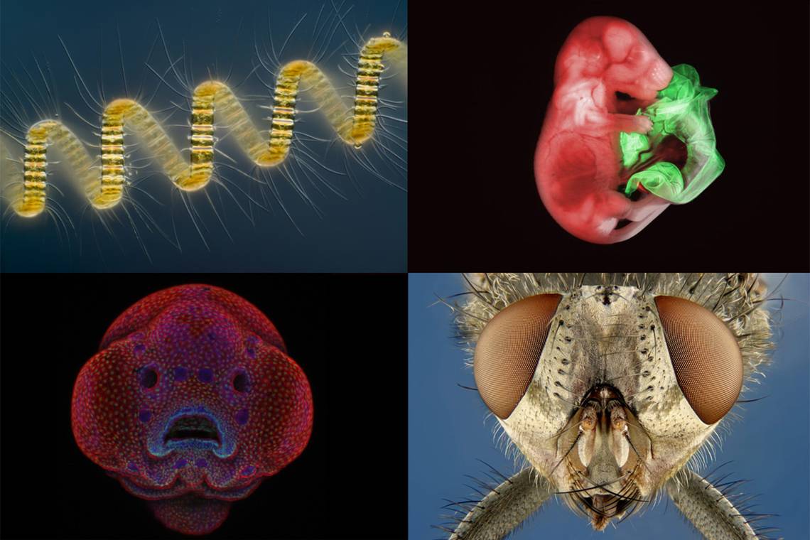
This image of a mosquito heart was the winning image selected from over 2,200 entries in 2010. The green dye shows up the musculature structure of the heart, while the blue dye shows the cells. A mosquito heart is a lot different to those in mammals and humans, it makes up the rear two-thirds of the circulatory system, which itself is made up of a tube that extends from the head to the tail. The heart is made up of a series of valves inside the tube, and helical coils of muscle that surround the tube and cause it expand and contract, which pumps blood around the body.
2011: Dr. Igor Siwanowicz, Max Planck Institute of Neurobiology, Martinsried, Germany

This 2011 winning image uses confocal imaging to show the intricate details that make up an invertebrate's inner structure.
- Subject Matter: Portrait of a Chrysopa sp. (green lacewing) larva
- Magnification: 20x
- Technique: Confocal
This 2011 winning image uses confocal imaging to show the intricate details that make up an invertebrate's inner structure. It's not a single image though, but rather a series of images that have been stitched together to show a much larger area. From it, we can see the various muscles inside the body that are used to operate the mouth.
2012: Dr. Jennifer L. Peters & Dr. Michael R. Taylor, St. Jude Children’s Research Hospital, Memphis, Tennessee, USA

- Subject Matter: The blood-brain barrier in a live zebrafish embryo
- Magnification: 20x
- Technique: Confocal
The blood-brain barrier is exactly what it says it is, a barrier that separates blood from the brain and extracellular fluid in the central nervous system (CNS). To create this image, Peters and Taylor developed a transgenic zebrafish (genetically modified) and then photographed using maximum intensity. The rainbow palette was used to not only produce a colourful image, but to provide spatial information.
2013: Wim van Egmond, Micropolitan Museum, Berkel en Rodenrijs, The Netherlands

Wim van Egmond had to employ an image stacking technique in order to show all the various structural elements of this particular plankton organism.
- Subject Matter: Chaetoceros debilis (marine diatom), a colonial plankton organism
- Magnification: 250x
- Technique: Differential Interference Contrast, Image Stacking
This 2013 winning image is another example of a diatom, one of the greatest producers of oxygen on earth. Wim van Egmond had to employ an image stacking technique (several different images layered on top of each other) in order to show all the various structural elements of this particular plankton organism. The DIC technique has resulted in a dark blue background that contrasts perfectly with the yellow colour of the specimen.
2014: Rogelio Moreno Gill, Panama, Panama

A second award-winning image of a rotifer. Rogelio Moreno Gill has photographed the interior of the mouth and the heart-shaped corona to win the award in 2014.
- Subject Matter: Rotifer showing the mouth interior and heart shaped corona
- Magnification: 40x
- Technique: Differential Interference Contrast
Another image of a rotifer won the Small World competition in 2014. This time, instead of showing one feeding, Rogelio Moreno Gill has photographed the interior of the mouth and the heart shaped corona. The corona is the crown that draws water into the mouth, which the rotifer then sifts through for any food.
2015: Ralph Grimm, Jimboomba, Queensland, Australia

- Subject Matter: Eye of a honey bee (Apis mellifera) covered in dandelion pollen
- Magnification: 120x
- Technique: Reflected Light
This extreme close up of the eye of a honey bee took Ralph Grimm over four hours to create. It clearly shows the intricate make up of a bee's eye, comprising hundreds of tiny hexagons. Grimm had to use reflective lighting to properly illuminate the sample, as direct incident light wouldn't have been able to reveal the same level of detail.
2016: Dr. Oscar Ruiz, The University of Texas MD Anderson Cancer Center, Houston, Texas, USA

Dr. Oscar Ruiz won the Small World competition with this confocal image of a four-day-old zebrafish embryo.
- Subject Matter: Four-day-old zebrafish embryo
- Magnification: 10x
- Technique: Confocal
Dr. Oscar Ruiz won the Small World competition with this confocal image of a four-day-old zebrafish embryo. Both its scientific and artistic value no doubt helped it to beat the competition. The confocal imaging technique, which captures multiple 2D images before stacking them to create a 3D image, helps to show the structural make up of zebrafish.
2017: Dr. Bram van den Broek et al, The Netherlands Cancer Institute, BioImaging Facility & Department of Cell Biology, Amsterdam, The Netherlands

The subject? Human skin, but with an excessive amount of keratin (shown in yellow), which is an important structural protein in the skin cell.
- Subject Matter: Immortalized human skin cells (HaCaT keratinocytes) expressing fluorescently tagged keratin
- Magnification: 40x (objective lens magnification)
- Technique: Confocal
One of the more recent winners of the Small World competition once again uses confocal imaging. The subject? Human skin, but with an excessive amount of keratin (shown in yellow), which is an important structural protein in the skin cell. Images such as this can help scientists look for various types of cancer, as reduced amounts of specific keratin can indicate an aggressive tumour.
2018: Yousef Al Habshi, Abu Dhabi, United Arab Emirates

- Subject Matter: The Eye of a beetle
- Magnification: 20x (objective lens magnification)
- Technique: Reflected Light
This incredible view almost looks too perfect to be real. An incredibly close-up view of a Metapocyrtus subquadrulifer beetle's eye. There have been many different entries into the photomicrograph competition over the years involving various different beetles and this might be one of the most impressive with wonderfully contrast between the black of the beetle's eye and the rest of its body.
2019: Teresa Zgoda, Teresa Kugler

This winner from 2019 is the result of a compilation of hundreds of images put together to form the final result. An amazing view of a turtle embryo.
- Subject Matter: A turtle embryo
- Magnification: 5x (objective lens magnification)
- Technique: Stereomicroscopy, Fluorescence
This winner from 2019 is the result of a compilation of hundreds of images put together to form the final result. The photographed turtle embryo was over an inch long and capturing the entire thing in a satisfying way was proving tricky for microscopy technician Teresa Zgoda and colleague Teresa Kugler.
The end result is a wonderful image which used a combination of fluorescence and stereomicroscopy to show off the beauty of the tiny turtle.
2020: Daniel Castranova, Dr. Brant M. Weinstein, Bakary Samasa, National Institutes of Health (NIH)

- Subject Matter: A zebrafish
- Magnification: 4x (objective lens magnification)
- Technique: Confocal
Taken using confocal microscopy and image-stacking, this incredible image shows not only the bones and scales but also the lymphatic vessels of a zebrafish. This image was not just a competition winner but also a scientific discovery as the photography revealed that zebrafish have lymphatic vessels inside their heads which wasn't thought to be the case with fish.
This knowledge could revolutionise future research into brain diseases such as Alzheimer's and thus made the image award worth on its own merit.
Daniel Castranova explained:
"The image is beautiful, but also shows how powerful the zebrafish can be as a model for the development of lymphatic vessels...Until now, we thought this type of lymphatic system only occurred in mammals. By studying them now, the scientific community can expedite a range of research and clinical innovations – everything from drug trials to cancer treatments. This is because fish are so much easier to raise and image than mammals."
2021: Jason Kirk, Baylor College of Medicine Houston, Texas, USA

- Subject Matter: Trichome and stomata of southern live oak leaf
- Magnification: 60X (Objective Lens Magnification)
- Technique: Image stacking
This incredible image was chosen as 2021's winner and shows an extreme close-up shot of a southern live oak leaf’s trichomes, stomata and vessels.
Jason Kirk is a professional imager and the core director for Baylor College of Medicine’s Optical Imaging & Vital Microscopy Core.
He used 200 individual images of the leaf stacked together to create the final imagery.
The white areas in the photo are trichomes, which are designed to help protect the plant against both insects and extreme weather conditions. The purple areas, meanwhile, show pores that regulate the flow of gases alongside the cyan coloured vessels that transport water throughout the leaf.
Incredible.
2022: Grigorii Timin and Dr. Michel Milinkovitch, University of Geneva

- Subject Matter: Madagascar giant day gecko
- Magnification: 63X (Objective Lens Magnification)
- Technique: Confocal
This one shows a pretty remarkable image of an embryonic hand of a Madagascar giant day gecko.
Here the imagers have used high-resolution microscopy and image-stitching to create a brilliant image of the gecko's hand.
Hundreds of images had to be stitched together to create this final product which won the first prize for 2022.